
Study Tests AI Screening for Coronary Disease Using Face and Tongue Images
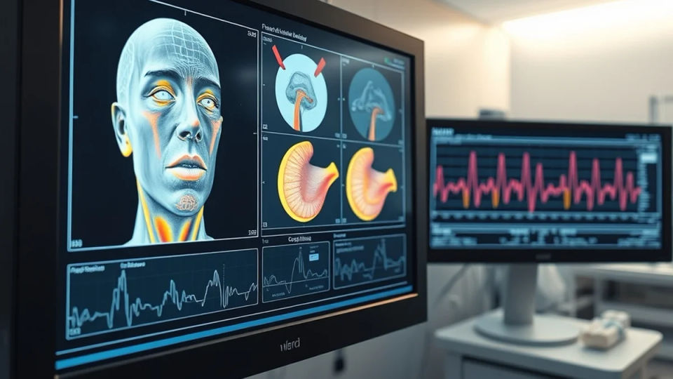
Researchers reported promising results for an experimental AI screening model using standardized photographs of the face and tongue in a case-control research cohort. Medical investigators enrolled 518 human subjects, including 200 patients clinically diagnosed with coronary heart disease alongside 318 healthy control participants. Each participant underwent standardized facial and oral photography using a purpose-built scanning apparatus, with resulting imagery parsed via multi-layer deep neural networks. The automated system evaluated lingual patterns and facial feature maps independently before synthesizing multi-modal predictions to calculate individual cardiovascular disease risk.
Deep Learning Facial and Lingual Feature Architecture
The algorithmic computer vision pipeline concentrated feature extraction filters across specific physiological zones of the facial surface and oral cavity. Automated neural attention centered on the core surface of the tongue, lingual fur coating thickness, circumoral skin textures, and bilateral cheek pigmentation. The study explored associations between image features and previously diagnosed disease; the visible patterns should not be used by the public as self-diagnostic signs. Coronary heart disease arises when primary arterial vessels supplying oxygenated blood to cardiac muscle tissue narrow due to lipid plaques, a condition termed atherosclerosis.
Clinical Cohort Testing and Blind Validation Accuracy
In a blinded validation test group comprising 104 individuals, the leading convolutional architecture attained an overall diagnostic classification accuracy of 98 percent. The neural diagnostic model successfully flagged all 40 coronary patients within the evaluation cohort, while generating two false-positive classifications among healthy participants. Investigators observed that combining facial imaging with tongue texture analysis delivered markedly superior diagnostic accuracy compared to examining either anatomical feature in isolation.
Methodological Limitations and False Positive Risks
The clinical trial, published in the peer-reviewed journal Scientific Reports, cautioned that testing established hospital patients against healthy controls differs from general population screening. Researchers highlighted that control group participants did not undergo invasive coronary angiography, meaning minor asymptomatic arterial lesions could not be completely excluded. Epidemiological specialists warned that deploying photographic screening tools across low-incidence general populations can yield substantial quantities of unneeded false-positive alarms.
Global Cardiovascular Disease Burden and Market Context
The international healthcare sector experiences continuous clinical urgency to expand diagnostic screening instruments capable of identifying early-stage coronary artery disease. Cardiology practices globally are shifting toward non-invasive diagnostic modalities that support early outpatient risk triage and accessible monitoring. Clinical adoption is propelled by escalating cardiovascular mortality linked to aging demographics, surging diabetes incidence, and adult obesity rates. Market research indicates commercial imaging hardware manufacturers are increasingly deploying artificial intelligence software to automate pattern recognition and improve diagnostic yield.
Why the striking scores need caution
The published study compared 200 patients with known coronary heart disease against 318 healthy control participants using standardized pictures and a machine-learning model. That case-control design is useful for exploring whether images contain predictive signals, but it is not the same as testing a device among people walking into a primary-care clinic with uncertain symptoms. The reported accuracy comes from a particular dataset, preprocessing procedure and evaluation split; it should not be extrapolated to all ages, skin tones, cameras or clinical populations.
A model can learn incidental differences between people recruited as patients and those recruited as controls. Independent, prospective studies at several hospitals would be needed to check whether it performs reliably when disease prevalence is lower and confounding factors vary. Performance measures such as sensitivity, specificity and false-positive rates would then determine whether an image-based tool actually helps clinical decisions.
This is experimental screening research, not an approved diagnosis.
A face or tongue photograph cannot replace clinical assessment, emergency care for possible heart-attack symptoms, blood tests or other established cardiac investigations. The paper's promising findings provide a basis for validation, not a reason for patients to self-diagnose.